AI in medical imaging: applications and challenges


AI in healthcare is more than a tool — it's transforming how we approach medical imaging. Imagine faster image analysis, earlier and more accurate detection of abnormalities, and treatment plans that are more precise and tailored to the patient. AI enables radiologists and specialists to see with greater clarity, revealing insights that might otherwise go unnoticed.
Our extensive experience in AI technology consultancy and healthcare software development has empowered organizations to tap into AI's immense potential in medical imaging — and we’re ready to share the discoveries.
The definition of AI in medical imaging
A 2023 report by the Royal College of Radiologists revealed a 30 percent shortage of radiology specialists in the UK, with projections indicating this could rise to 40 percent by 2028 if no further action is taken. And this critical shortage isn't confined to the UK. Similar situations are evident globally — in Australia, New Zealand, and Africa, where there are as few as 1.5 radiologists per million people.
In the US, for instance, the American College of Radiology (ACR) job board lists thousands of open radiologist positions, highlighting the widespread demand and the global challenge of meeting this need.
As the demand for skilled radiologists continues to outpace supply, AI in medical imaging is emerging as a vital solution. It’s a technology that leverages deep learning algorithms and machine learning techniques to analyze visual representations of body organs — such as X-rays, CT scans, fluoroscopy, and MRI scans — enhancing diagnostic accuracy and efficiency.
AI in medical imaging can detect even the smallest changes or abnormalities in images that might be invisible to the human eye, making it an invaluable tool for doctors to diagnose diseases accurately. Even more importantly, AI is making significant strides in diagnosing advanced diseases like lung cancer, identifying conditions requiring treatment, and predicting long-term survival factors.
AI in medical imaging market: key stats
With 15 years in the industry, our healthtech delivery manager, Eugene Kruglik, has witnessed the industry's evolution firsthand. "Breakthroughs in AI, machine learning, big data, wearables, and genomics signal we're on the cusp of a healthcare revolution. Those who invest in AI today are positioning themselves as leaders in this transformation."
The statistics back up Eugene's prognosis: more and more medical professionals will inject AI into their workflows.
Market size
- Globally, AI in the medical imaging market was estimated at $0.98B in 2023 and is expected to soar to $14.27B by 2032, with a registered CAGR of 33.1 percent during the forecast period from 2023 to 2032.
- In the US, AI in the medical imaging market was valued at $170.56M in 2023 and is projected to expand at a compound annual growth rate of 29.16 percent from 2024 to 2033, reaching more than $2B.
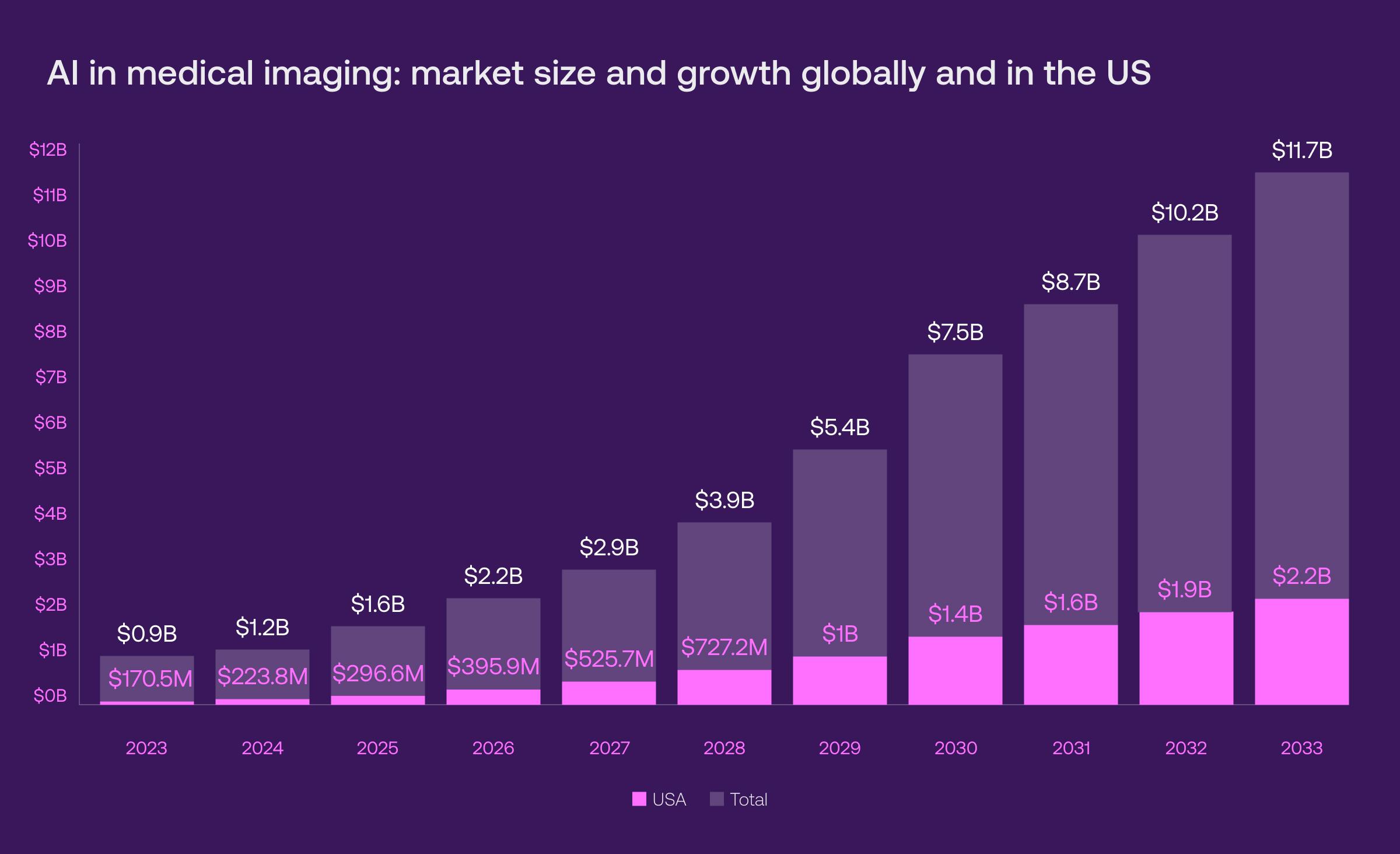
Source: precedenceresearch.com
-
The deep learning market led the pack in 2022, with a 58.8 percent share of its revenue among other technologies. However, this trend soon shifted, with the NLP segment coming to the stage from 2023 to 2032 with its fastest CAGR.
-
Neurology (20.95 percent of revenue share in 2022) and breast screening segments (35.6 percent) are the leaders in the market by application criteria.
-
The CT scan segment led in 2022 with a revenue share of 37.4 percent. The X-ray segment follows, with the strongest CAGR of 35.4 percent from 2023 to 2032.
Capabilities of AI in medical imaging
Computer-aided diagnosis (CAD)
AI CAD systems are designed to assist medical professionals in making more accurate and timely diagnoses. These systems are trained on vast medical image databases, including examples of various diseases and abnormalities. Once trained, AI algorithms can automatically detect and highlight potential areas of concern in images, often at early stages, enabling more effective treatment or timely intervention.
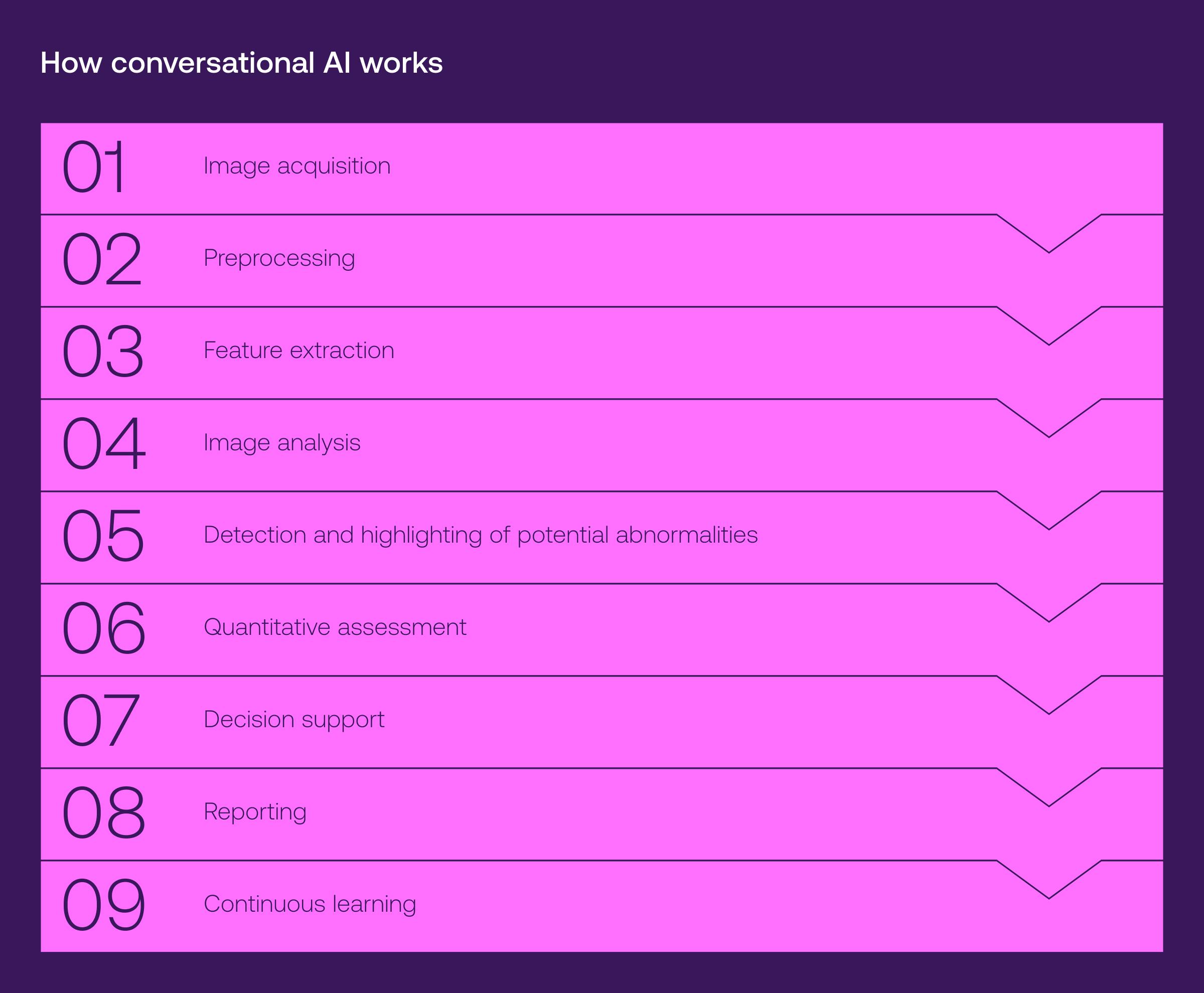
CAD modalities
AI CAD systems are highly adaptable across different medical imaging modalities. Whether picking up anomalies in a chest X-ray, detecting lesions in a mammogram, or analyzing structural details in an MRI scan, AI CAD is tailored to interpret a wide range of medical images.
Image segmentation
AI segmentation medical imaging is a computer vision technique that identifies specific parts of a medical image based on various criteria. Its main goal is to isolate areas of interest within the image, which is crucial for object recognition, scene understanding, and medical image analysis.
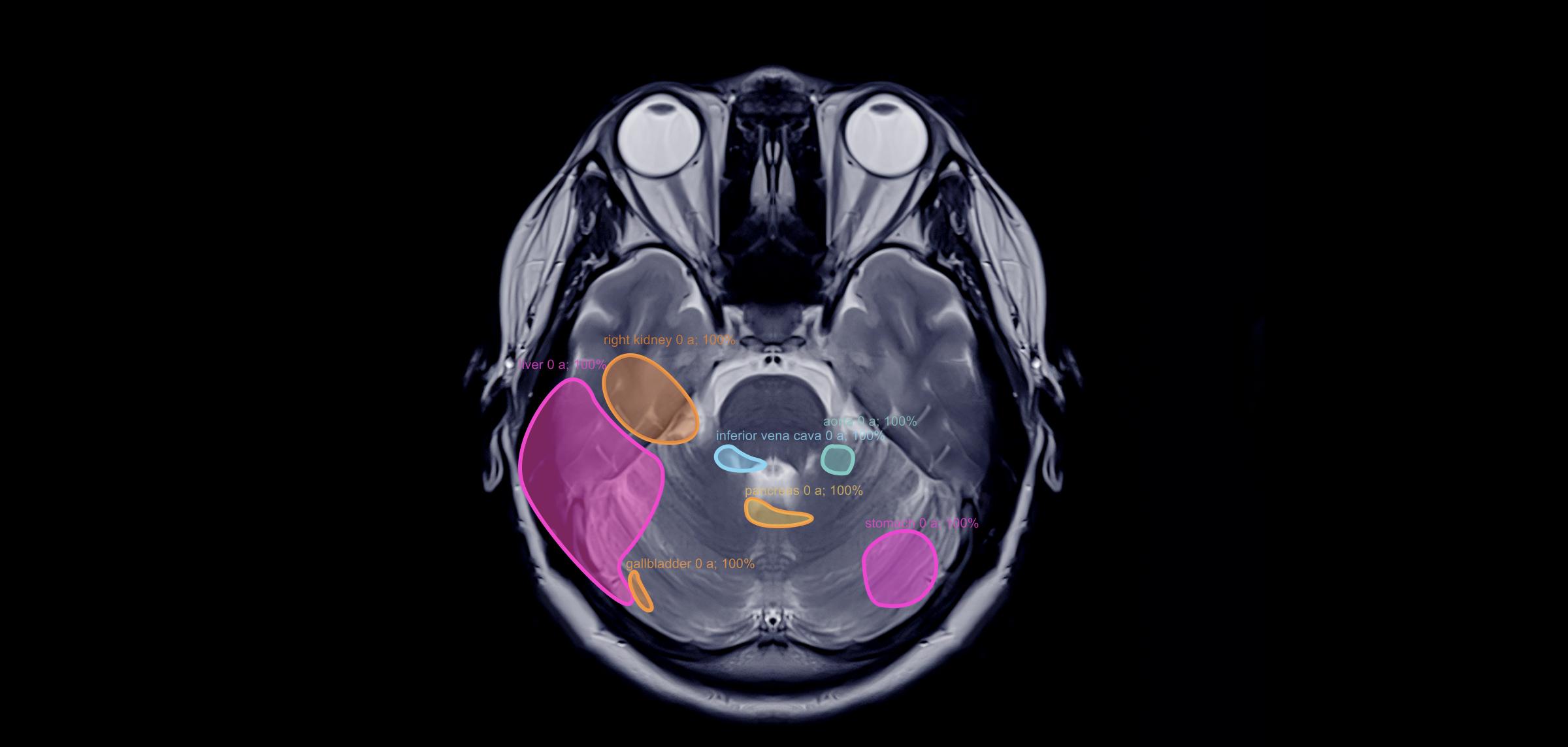
There are two types of image segmentation:
Semantic segmentation
When a detailed understanding of different objects or regions in an image is needed, semantic segmentation comes into play. This technique classifies each pixel in an image into a specific class or category, providing a precise analysis of the image's components.
AI-based algorithms help identify specific structures within medical images and provide precise quantitative analysis of areas of interest.
Instance segmentation
Unlike semantic segmentation, instance segmentation assigns a unique tag to each distinct object in the image, rather than grouping all pixels of a particular class. This approach is essential when multiple objects of the same class appear in close proximity, allowing for precise differentiation between them.
Image segmentation usage
Image segmentation is widely used to analyze X-rays, CT scans, MRI scans, and ultrasound images. For instance, in a chest CT scan, segmentation helps identify and outline the lungs, heart, and other vital structures, aiding in detecting abnormalities like tumors or lesions.
It can also differentiate between structures within a single organ and is especially valuable for analyzing smaller objects such as tissue samples or cells.
Image segmentation technologies
Several major image segmentation techniques are commonly used, including thresholding, edge detection, region-based segmentation, clustering methods, and neural network-based segmentation. The latter leverages deep learning models, particularly convolutional neural networks (CNNs), with methods like fully convolutional networks (FCNs), U-Net, and Mask R-CNN.
Medical imaging workflow optimization
“Optimizing workflows is always at the top of our clients' lists. When they come to us, they look for EMR/EHR systems, automated reporting, and patient portal development," says Pavel Nekrasov, ML Engineering Manager at Vention. "With the AI power, no one wants to spend unnecessary time on administrative tasks, whether a surgeon or a radiologist.”
Here’s how AI in medical imaging is transforming healthcare, streamlining workflows, enhancing efficiency, and delivering spot-on diagnoses and treatments:
Automated protocoling
AI algorithms automatically choose the right imaging protocols based on the patient’s specific clinical needs, which not only reduces the manual work involved, but also ensures that imaging procedures are standardized and efficient.
Image reconstruction
Leveraging deep learning algorithms, lower-dose or quicker scans are transformed into high-quality images. Extra scans are no longer necessary, saving patients time, effort, and money.
Workflow prioritization and triage
AI flags potential abnormalities, enabling radiologists to prioritize cases requiring immediate attention. This process ensures that critical patients receive urgent care when it’s most needed.
Automated reporting
AI algorithms trained on medical documentation can automatically generate structured reports based on imaging results. By ensuring consistency across all documentation, more time is freed up for direct patient care.
Image quality assessment
AI algorithms make it quick and easy to assess the quality of medical images, preventing artifacts or suboptimal acquisition parameters from entering the diagnostic workflow.
Integration with electronic health records (EHR)
When integrated with AI, EHR systems can automatically update all imaging data in a patient's history, improving continuity of care and fostering better collaboration among healthcare professionals.
Quantitative analysis
AI-driven quantitative medical imaging analysis revolutionizes how clinicians interpret and utilize imaging data. By automatically measuring and analyzing features such as tumor size, tissue density, and organ volume, AI tools provide precise, objective insights that enhance diagnostic accuracy and treatment planning.
Automated measurements
Advanced AI algorithms automatically measure key parameters from imaging data, such as lesion and tumor size, organ volume, and vessel diameters, as well as track their changes over time. Automation reduces the need for manual measurements, minimizing human error and freeing up valuable time for radiologists to focus on patient care.
Functional imaging analysis
AI not only examines the structure of organs and tissues, but also analyzes their function through dynamic contrast-enhanced MRI and CT perfusion scans. It identifies blood flow, perfusion, tissue enhancement kinetics, and functional connectivity patterns in the brain, which is crucial for neurological conditions.
Quantitative biomarker extraction
Radiomics, which extracts extensive features from medical images using data-characterization algorithms, reveals tumor patterns and characteristics not visible to the naked eye.
AI is key in identifying and extracting quantitative biomarkers from medical images, offering valuable insights for diagnosing diseases, predicting outcomes, and assessing treatment efficiency.
Texture feature extraction
AI is used to analyze the texture and spatial patterns of tissues to characterize diverse types or identify abnormalities. It also assists in quantifying the characteristics of atherosclerotic plaques — plaque volume, composition, and vulnerability — by analyzing cardiovascular imaging.
Disease progression monitoring
By analyzing imaging data over time, AI algorithms can detect subtle changes in disease states that the human eye might miss. This allows for early intervention, more accurate tracking of disease evolution, and better-informed decisions about treatment adjustments.
AI medical imaging software solutions
Explore AI-powered medical imaging software that delivers accurate diagnoses, streamlined workflows, and better patient outcomes.
Tempus Radiology delivers AI-driven insights from medical images, offering automated lesion segmentation and measurement, longitudinal tracking for lesion response, and automated reporting. These tools streamline diagnosis and treatment response and integrate effortlessly with patient records.

Nanox makes medical imaging technology and solutions more accessible through products like Nanox ARC, a cost-effective digital X-ray device, and Nanox Cloud, a cloud-based platform for image storage, sharing, and AI-powered analysis. Together, they provide accessible, high-quality medical imaging and remote diagnostics.

Infervision specializes in AI medical solutions that support the entire patient treatment cycle, from disease screening and diagnosis to intervention, patient management, and medical research. They focus on developing advanced algorithms to assist radiologists in detecting and diagnosing conditions like lung cancer, hemorrhage, stroke, and pneumonia.

Qure.AI leverages algorithms trained on 4.4 million scans to analyze various medical images, including X-rays, CT scans, and MRIs. Their tools are designed to detect abnormalities, facilitate early diagnosis of conditions such as lung disease, head trauma, and tuberculosis, and automatically generate reports, highlight areas of concern, and integrate seamlessly with existing healthcare systems.

Subtle Medical offers AI-powered solutions like SubtlePET and SubtleMR, which enhance PET and MRI image quality, enabling faster scans and reduced radiation. Their tools integrate with existing systems to optimize imaging workflows (save up to 25 percent of the original scan time) and improve patient care with a shorter exam time.

AI imaging healthcare: proven results and outcomes
Medical imaging and AI have statistically proven to be highly effective across a range of use cases among researchers:
LVO strokes detection
In a study conducted by Viz, a medical imaging company, the Viz.AI LVO platform — which analyzes brain CT angiography (CTA) images for suspected acute ischemic stroke (AIS) and automatically alerts clinicians to potential large vessel occlusions (LVO) — demonstrated impressive results.
The research confirmed the high reliability of Viz.AI LVO, with a sensitivity of 96.3 percent and specificity of 93.8 percent, making it a trusted tool for neuroradiologists in emergency settings.
Hemorrhage recognition
A study at Mount Sinai Hospital found that Gauss Surgical’s Triton system significantly improved AI recognition of postpartum hemorrhage in medical imaging, leading to earlier interventions and substantial cost savings.
By accurately tracking blood loss in real-time during over 7,600 deliveries, Triton improved hemorrhage detection and cut down on unnecessary transfusions and lab tests, saving the hospital more than $209K annually.
Radiology abnormalities detection
Aidoc tackles the challenge of rising radiologist workloads with AI-powered decision-support software that efficiently analyzes CT scans. A 2023 study published in RSNA Journals assessed Aidoc's software for identifying incidental pulmonary emboli (iPE) in cancer patients' chest CT scans.
The study reviewed 11,736 scans and found that AI reduced the occurrence of iPE from 1.3 -1.4 percent to 1 percent, demonstrating 91.6 percent sensitivity, 99.7 percent specificity, and 99.9 percent negative predictive value, which proved its effectiveness in routine clinical practice.
Carcinoma classification
PathAI transforms pathology with advanced machine learning algorithms, particularly in analyzing ovarian cancer tissue images. Trained on vast datasets, PathAI’s technology can accurately classify various tissue components, such as cancer cells, fibroblasts, and inflammatory cells, matching the precision of pathologists.
The study also uncovered a link between the diversity of cancer cell nuclei and patient outcomes, including overall survival. This finding suggests that PathAI’s model could be instrumental in classifying ovarian cancer subtypes and studying patient responses to targeted therapies, potentially influencing future treatment strategies.
Gestational age estimation
Butterfly iQ is a groundbreaking handheld ultrasound device that uses AI to improve image acquisition and interpretation. In 2021, researchers trained a neural network on scans from 4,695 pregnant women in North Carolina and Zambia to estimate gestational age.
The AI showed incredible results: it outperformed traditional fetal biometry methods, offering a promising solution for areas with limited access to trained professionals. This innovation highlights AI's potential to enhance diagnostic accuracy, especially in remote areas with restricted medical resources.
Need AI healthcare technology experts for your project?
Our developers are ready to assess your needs, deliver AI-powered solutions, and elevate your systems.
Challenges of AI-based medical imaging and practical solutions
By recognizing the current challenges in AI for medical imaging, specialists can work through them to improve diagnostic accuracy and make the diagnostic process even more reliable and secure.
Data quality
|
Challenges |
Practical solutions |
|
Need for large and diverse datasets: AI models require extensive and varied datasets for effective training.
Non-representative training data: training data that doesn't represent the entire population can lead to inaccuracies, particularly in underrepresented groups.
Labor-intensive labeling: labeling medical images for training datasets can be time-consuming and prone to errors.
Impact of inaccurate or biased labels: inaccurate or biased labeling can negatively affect the performance and reliability of AI models. |
Include diverse populations: this improves data quality and ensures balanced representation across different demographics, ethnicities, and clinical conditions.
Use data augmentation: apply transformations like rotations, flips, and contrast changes to existing images to generate new training samples.
Leverage adversarial networks: help the model learn from data and potential mistakes, increasing its robustness.
Implement explainable AI (XAI) and bias detection tools: identify where mistakes might occur and guide corrective actions. |
Interoperability and integration
|
Challenges |
Practical solutions |
|
Integration with existing healthcare systems: incorporating AI solutions into current electronic health records (EHRs) and imaging infrastructure can be complex.
Lack of standardized data formats and protocols: inconsistent data formats and protocols across healthcare institutions hinder the effective sharing and integration of medical imaging data. |
Adopt industry standards: adhere to widely accepted medical imaging data standards like DICOM to ensure smooth integration of AI applications.
Utilize open APIs: facilitate the exchange of information between AI solutions and existing healthcare infrastructure, promoting interoperability.
Leverage health information exchange platforms: enable the secure exchange of patient information among healthcare organizations and serve as intermediaries for data sharing between AI applications.
Collaborate with healthcare IT vendors: align AI applications with existing healthcare technologies and standards by working with reliable healthcare IT vendors and get a consultancy on interoperability in healthcare systems. |
Clinical validation and real-world performance
|
Challenges |
Practical solutions |
|
Need for rigorous clinical validation: many AI algorithms in medical imaging require thorough validation to prove their effectiveness and reliability in real-world healthcare settings.
Trust issues due to lack of robust validation: gaining trust among healthcare professionals can be challenging without solid clinical validation.
Generalization challenges across diverse populations: AI models trained on specific datasets may struggle to perform well across different populations, conditions, or imaging devices.
Impact on real-world performance: limited generalization can negatively affect the AI's effectiveness in actual clinical practice. |
Conduct diverse clinical trials: including a wide range of patient populations and real-world clinical settings helps assess the algorithm's effectiveness in various conditions.
Engage clinicians: involve radiologists, pathologists, and other relevant specialists to improve clinical workflows, address challenges, and evaluate AI's impact on patient care.
Seek independent validation: obtain third-party validation to ensure the model's generalizability and performance across different contexts.
Benchmark AI performance: compare AI performance against established standards to assess its contribution directly and ensure it adds value beyond existing practices.
Implement transparent reporting standards: use standards like Consolidated Standards of Reporting Trials to enhance the reproducibility and credibility of study results. |
Interpretability and explainability
|
Challenges |
Practical solutions |
|
The perception of deep learning models as "black boxes": neural networks are often seen as opaque due to their complex structures.
Need for expandable AI in medical imaging: understanding how AI arrives at specific diagnoses or recommendations is crucial for building trust among healthcare professionals and patients. |
Use LIME and SHAP: employ techniques like LIME (local interpretable model-agnostic explanations) and SHAP (Shapley additive explanations) to generate post-hoc explanations for black-box models.
Highlight regions of interest: utilize algorithms that identify key areas in images, helping clinicians understand which parts of the image are driving the AI's predictions.
Incorporate attention mechanisms: for deep learning models, use attention mechanisms to emphasize important features or regions in the input data that influence the model's decisions.
Develop interactive visual tools: provide tools that allow clinicians to manipulate input features and observe how predictions change in response.
Provide uncertain information: offer details about the uncertainty associated with the model's predictions to help clinicians gauge the reliability of AI-generated results. |
Cybersecurity and privacy
|
Challenges |
Practical solutions |
|
Large volumes of sensitive patient data: AI applications in healthcare generate and process significant amounts of sensitive medical information.
Importance of robust cybersecurity measures: ensuring strong protections against unauthorized access, data breaches, and malicious attacks is crucial.
Patient concerns about privacy and security: patients may worry about the safety of their medical information when AI is used for data analysis and storage. |
Anonymize and de-identify data: remove personally identifiable information to reduce the risk of patient re-identification.
Implement role-based access control (RBAC) and multi-factor authentication (MFA): enhance security by requiring users to provide multiple forms of identification for system access.
Use secure communication protocols and data encryption: protect systems from unauthorized access, eavesdropping, and data tampering.
Ensure compliance with regulations: adhere to healthcare and data protection regulations like HIPAA or GDPR to establish a framework for safeguarding patient privacy. |
AI medical imaging software development at Vention
Overcoming AI's challenges and limitations in medical imaging requires the right IT partner—one with a strong portfolio and extensive experience to ensure your project's success.
At Vention, we’re dedicated to creating transparent, secure, and effective AI solutions that empower medical professionals to analyze images more efficiently, reduce administrative burdens, and keep the focus where it truly belongs — on patient care.
For example, our team collaborated with Imagen Technologies, a company specializing in accurate, accessible, and affordable AI-driven medical image interpretation, to enhance their computer vision system for diagnostic imaging. “Throughout our partnership, we also rolled out new web tools, scripts, and admin interfaces that enabled Imagen’s research team to manage data labeling independently, without relying on engineers. This streamlined radiology workflows and accelerated condition detection,” says Eugene Kruglik, Healthcare Development Expert at Vention.
Recognizing the importance of security, especially amidst numerous healthcare cybersecurity challenges, we also prioritize stringent security standards in all our projects. “We help you craft solutions that fully comply with HIPAA, GDPR, PCI DSS, and other standards, and we’re confident that our work will continue to pave the way for the next generation of healthcare innovators,” notes Pavel Nekrasov.
So, Venton is your go-to if you're looking to transform your organization into a revolutionary force in healthcare. We handle every aspect of the development process, giving you the peace of mind to focus on what truly matters — delivering exceptional patient outcomes.
Seeking assistance with your AI project?
We’ve got the unique talent you need for AI development.


